
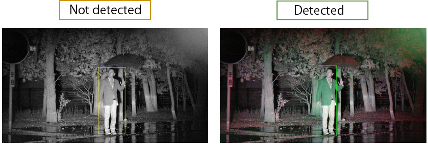
Improved detection capabilities in surveillance
Who was it?..
The guy in the BLUE jacket!
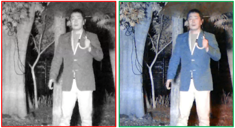
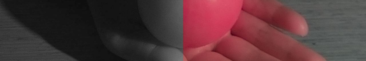
Color Night Vision
Detection
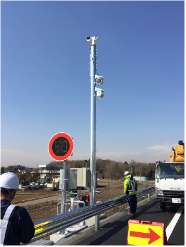
Nanolux camera in service
on the highway, since Feb 26, 2017
- Monitoring 500m away on the both sides.
- The construction cost is 1/8 of the conventional camera and visible illuminators.
Color night vision : NEXCO
Left: 4 fps conventional camera Right: Nanolux camera 30 fps
3x Accuracy
(machine learning)
| Color IR | Mono IR | |
|---|---|---|
| Correct recognition | 67 | 19 |
| Error recognition | 34 | 173 |
Evaluation by Soft Solution Co. , a third party vendor
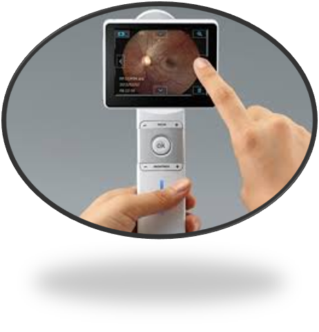
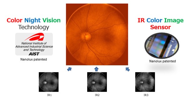
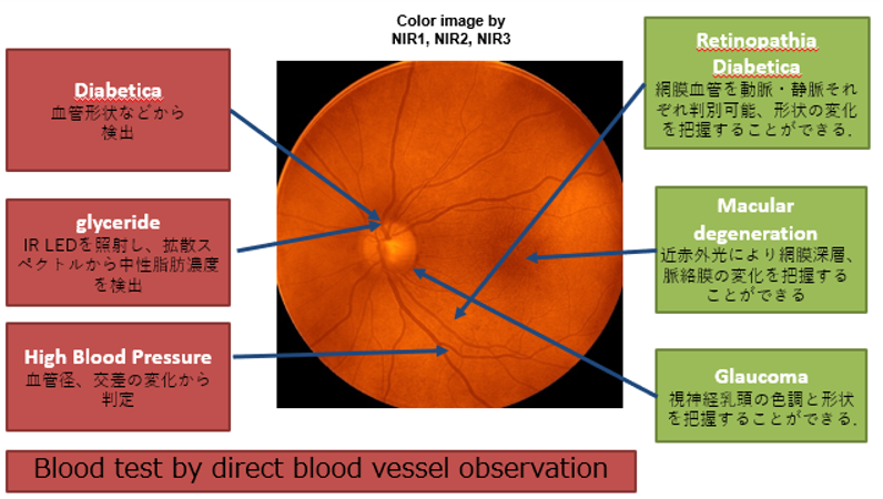
Glare-free color IR fundus camera
Smartphone Fundus Camera
Develop a fundus camera in the mobile-form factor to excel the smart health agenda
Advantages
- Glare-free
- Non-invasive blood test
- Color IR
- Movie
- No miosis
- AI diagnosis
What can be inspected
- Glucose
- Blood fat
- Arteriosclerosis
- Eye diseases, etc
Source:Nara Institute of science and technology
Development of a practical machine for a “Glare-Free” near-infrared color fundus camera, and started verification at Osaka University Hospital

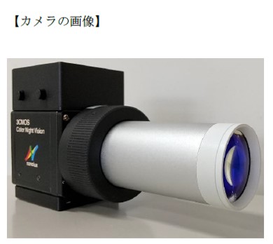
Nanolux Co., Ltd. (Headquarters: Ushiku City, Ibaraki Prefecture, CEO: Motoshi Sobue) which develops, designs, and manufactures “near-infrared color night vision technology” that enables color photography even in the dark and Nara Institute of Science and Technology (Nara Prefecture, Ikoma City, President: Naokazu Yokoya) Professor Atsushi Ota of the Optical Functional Element Science Laboratory, Graduate School of Science and Technology, have succeeded in developing a practical machine that can be used for verification in the medical field of a “Glare-Free fundus camera” that does not require visible illumination light but shoot with near-infrared light as the light source. In addition, Osaka University Graduate School of Medicine Eye Science (Professor: Koji Nishida) and Osaka University affiliated Hospital AI Medical Center (Specially Appointed Professor (Full-time): Ryo Kawasaki), who are conducting joint research with Nanolux Co., Ltd., have started verification at the University Hospital (hereinafter referred to as “Osaka University Hospital”).
A normal fundus camera is very dazzling because it uses a visible light flash to shoot the fundus, and with non-mydriasis, the pupils are miotic in the first shot, making repeated shooting difficult and shooting for children. Since this camera uses only near-infrared light and does not use flash light, it is less invasive than conventional fundus cameras, is safer, and has wide-ranging and detailed observation of the structure including blood vessels of the eyeball with non-mydriasis. Furthermore, by making it possible to easily capture fundus images with less burden on the patient, it is expected that fundus photography will become easier and contribute to early detection of lifestyle-related diseases such as hypertension as well as eye diseases. The newly developed ” Glare-Free fundus camera” has achieved miniaturization and high operability by combining Nanolux’s near-infrared color fundus camera NLX-FD001 with a cylindrical lens integrated with near-infrared illumination. In addition, the purpose of verification at Osaka University Hospital is to observe major anatomical sites and normal & abnormal findings with actual patients using a near-infrared fundus camera to confirm the usability of doctors. Therefore, it would be extremely clinically meaningful if it could be examined with a near-infrared fundus camera with minimal invasiveness. So far, more than 10 photographs have been taken at hospital sites, and it has been confirmed that there is no problem in practical operation. This research verification is scheduled to continue until March 2021.This camera development was carried out with the support of the Japan Science and Technology Agency (JST) Strategic Creative Research Promotion Project ACCEL (JPMJAC1601).
Nanolux Fundus Camera NLX-FD001(PDF)
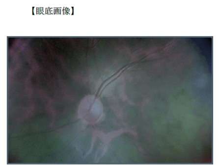
Accumulated fundus image
Concept phase been completed
- The sample camera was developed.
- The color IR fundus images were taken successfully.
- It’s a non-flashy and selfie.
- A medical doctor endorsed the effectiveness of color IR images.
- The outcome was reported and recognized by the academic magazine.
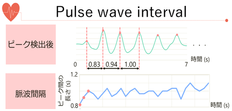
Detect vital information from pulse wave data
Detection of stress index from heartbeat interval
Pulse wave detection of driver/passenger
abnormality

To be developed in future projects
POC: Human Sensing
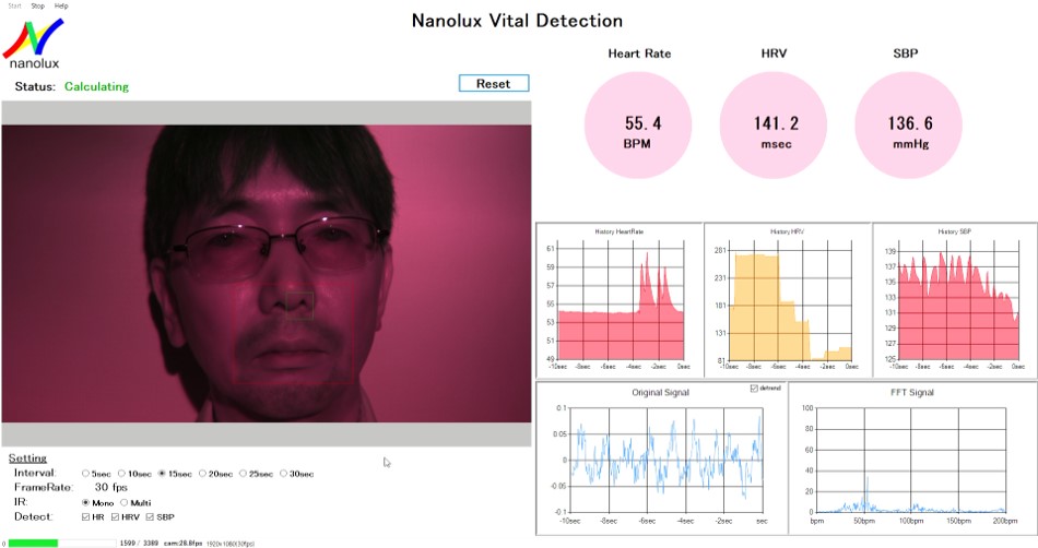
- Pulse wave can be measured only by IR.
- Noninvasive & No-phycial contact.
- 24×7 monitoring without annoying the target.
- Best fit to automotive and patient care.